Lessons and Practice Strips
ECG Practice Strips
Lesson #1: Rhythm Analysis Method
Intro
The five steps of rhythm analysis will be followed when analyzing any rhythm strip. Analyze each step in the following order.
- Rhythm Regularity
- Heart Rate
- P wave morphology
- P R interval or PRi
- QRS complex duration and morphology
Step 1
Rhythm Regularity
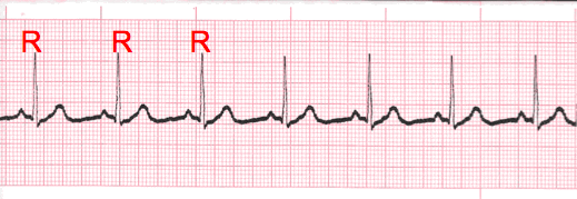
- Carefully measure from the tip of one R wave to the next, from the beginning to the end of the tracing.
- A rhythm is considered “regular or constant” when the distance apart is either the same or varies by 1 ½ small boxes or less from one R wave to the next R wave.
Step 2
Heart Rate Regular (Constant) Rhythms

- The heart rate determination technique used will be the 1500 technique.
- Starting at the beginning of the tracing through the end, measure from one R wave to the next R wave (ventricular assessment), then P wave to P wave (atrial assessment), then count the number of small boxes between each and divide that number into 1500. This technique will give you the most accurate heart rate when analyzing regular heart rhythms. You may include ½ of a small box i.e. 1500/37.5 = 40 bpm (don’t forget to round up or down if a portion of a beat is included in the answer).
Step 2 (Cont.)
Heart Rate - Irregular Rhythms

- If the rhythm varies by two small boxes or more, the rhythm is considered “irregular”.
- The heart rate determination technique used for irregular rhythms will be the “six-second technique”.
- Simply count the number of cardiac complexes in six seconds and multiply by ten.
Step 3
P wave Morphology (shape)

- The lead most commonly referenced in cardiac monitoring is lead II.
- In this training module, lead two will specifically be referenced unless otherwise specified.
- The P wave in lead II in a normal heart is typically rounded and upright in appearance.
- Changes in shape must be reported. This can be an indicator that the locus of stimulation is changing or the pathway taken is changing.
- P waves may come in a variety of morphologies i.e. rounded and upright, peaked, flattened, notched, biphasic(pictured), inverted and even buried or absent!
- Remember to describe the shape. This can be very important to the physician when diagnosing the patient.
Step 4
PR interval (PRi)
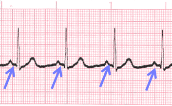 Constant PR Interval
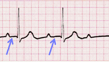 Variable PR Interval
- Measurement of the PR interval reflects the amount of time from the beginning of atrial depolarization to the beginning of ventricular depolarization.
- Plainly stated, this measurement is from the beginning of the P wave to the beginning of the QRS complex.
- The normal range for PR interval is: 0.12 – 0.20 seconds (3 to 5 small boxes)
- It is important that you measure each PR interval on the rhythm strip.
- Some tracings do not have the same PRi measurement from one cardiac complex to the next. Sometimes there is a prolonging pattern, sometimes not.
- If the PR intervals are variable, report them as variable, but note if a pattern is present or not.
Step 5
QRS complex
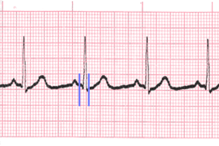
- QRS represents ventricular depolarization.
- It is very important to analyze each QRS complex on the tracing and report the duration measurement and describe the shape (including any changes in shape).
- As discussed in step 3, when referring to P waves, remember changes in the shape of the waveform can indicate the locus of stimulation has changed or a different conduction pathway was followed. It is no different when analyzing the QRS complex. The difference is that in step 3, we were looking at atrial activity. Now we are looking at ventricular activity.
- Measure from the beginning to the end of ventricular depolarization.
- The normal duration of the QRS complex is: 0.06 – 0.10 second
Lesson #2: Interpretation 313
Introduction
- The previous slides presented the five-steps of rhythm analysis. These five steps must be followed regardless of how simple of complex the tracing is you are reviewing.
- The information gathered in these steps are telling a story.
- The title of that story is the interpretation.
Sinus Rhythm Types
The dysrhythmias in this category occur as a result of influences on the Sinoatrial (SA) node. Rhythms in this category will share similarities in a normal appearing P wave, the PR interval will measure in the “normal range” of 0.12 – 0.20 second, and the QRS typically will measure in the “normal range” of 0.06 – 0.10 second. For the most part, dysrhythmias in this category either effect the rate, rhythm regularity or both within a particular tracing. We will be discussing the following complexes and rhythms:
- Normal Sinus Rhythm
- Sinus Bradycardia
- Sinus Tachycardia
- Sinus Dysrhythmia (Arrhythmia)
- Sinus Arrest
- Sinus Exit Block
Lesson #3: Normal Sinus Rhythm
Introduction

- Also known as Sinus Rhythm is the only rhythm when each of the five steps of rhythm analysis are “normal”.
- All other rhythms you will analyze will have at least one of the 5-steps presenting an abnormality.
- This rhythm will be regular, in a heart rate range between 60 – 100 bpm, P waves are upright and uniform in appearance (in Lead II), with a P wave for each QRS complex, the PR interval will measure in the normal range of 0.12 – 0.20 second (and measure the same each time), and the QRS complex morphology will be similar beat-to-beat and measure between 0.06 – 0.10 second (and measure the same each time).
Practice Strip

Analyze this tracing.
Show Answer
- Rhythm: Regular
- Rate: 75 (20 small boxes)
- P wave: Upright & uniform
- PR interval: 0.16 second
- QRS: 0.08 second
- Normal Sinus Rhythm
Lesson #4: Sinus Bradycardia
Description

- Rhythms are often named according to the origin of the electrical activity in the heart or the structure where the problem is occurring.
- Sinus Rhythms are aptly named due to the locus of stimulation being the SA (sinoatrial) node.
- With Sinus Bradycardia the locus of stimulation is the same as normal sinus rhythm, just now the rate will be less than 60 bpm. Recall that “brady” means slow.
- The only difference between sinus bradycardia and normal sinus rhythm is the rate. All other steps in the rhythm analysis are “normal”.
Practice Strip

- Analyze this tracing using the five steps of rhythm analysis.
- Compare your answers with the answers on the next slide.
Show Answer
- Rhythm: Regular
- Rate: 56 (27 small boxes)
- P wave: Upright and Uniform
- PR interval: 0.18 sec
- QRS: 0.10 sec
- Interpretation: Sinus Bradycardia
Lesson #5: Sinus Tachycardia
Description
- Sinus Tachycardia occurs when the rate of electrical impulse formation occurs at a rate exceeding 100 bpm.
- This can occur for a number of different reasons i.e. diet, stress, illness, response to physical exertion etc.
- The only difference between Normal Sinus Rhythm and Sinus Tachycardia (Sinus Tach) is the rate exceeds 100 bpm. All other steps of rhythm analysis will be “normal.
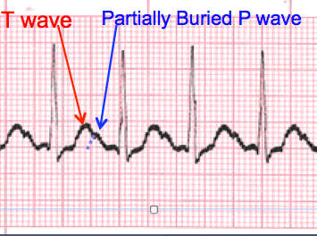
- An additional challenge that will present as rhythm rates accelerate is that the cardiac complexes will come closer together. This can result in the P wave becoming partially or completely buried within the T wave of the previous cardiac complex.
- The result of a partially buried P waves means you are unable to establish the beginning of atrial depolarization. Meaning you will be unable to measure and report the PR interval. The only way it will be possible is if the physician instructs you to increase the machine printing speed (remember interval times will double at 50 mm/sec).
Practice Strip

Analyze this tracing using the five steps of rhythm analysis.
Show Answer
- Rhythm: Regular
- Rate: 125
- P wave: upright, partially buried
- PR interval: unable to determine
- QRS: 0.08 sec
- Interpretation: Sinus Tachycardia
Lesson #6: Sinus Dysrhythmia (Arrhythmia)
Description

- Sinus Dysrhythmia often occurs as a normal variant. It is commonly seen in young healthy people and athletes. It is frequently related to breathing and pressure on the vagus nerve. As the patient inhales and the lungs expand, pressure is applied to the vagus nerve which causes a parasympathetic response and a decrease in heart rate.
- Sinus Dysrhythmia ECGs may also occur as a result of medication effects or diseases such as multiple sclerosis.
- This dysrhythmia often requires no treatment, but may require medication or therapy such as a pacemaker to regulate the heart rate if the ventricular response becomes too slow.
- Sinus Dysrhythmia closely resembles Normal Sinus Rhythm with the only distinction being the intervals from one cardiac complex to the next are changing as influenced by the patients respiratory pattern.
- Note the changing R to R Intervals.
Sinus Dysrhythmia EKG Practice Strip

Analyze this tracing using the five steps of rhythm analysis.
Show Answer
- Rhythm: Irregular
- Rate: 60 bpm
- P wave: upright and uniform
- PR interval: 0.16 sec
- QRS: 0.10 sec
- Interpretation: Sinus Dysrhythmia
Lesson #7: Sinus Arrest
Description
- Sinus Arrest occurs when there is a sudden absence of electrical activity initiated by the SA node. This results in a pause in the electrical activity seen on the tracing. Remember, no electrical activity = no depolarization and contraction. Hence, a drop in blood pressure.
- The longer the pause, the further the blood pressure will drop.
- A pause of six-seconds is considered a medical emergency and emergency procedures must be initiated.
- The ECG sinus arrest rhythm typically will demonstrate constant R to R intervals prior to and following the pause. This pause results in the rhythm tracing presenting as irregular.
Analysis

- It is important to measure the duration of the pause and report this information along with the frequency of the pauses if there are more than one.
- The pause is measured by placing your caliper tips on the last R wave just prior to the pause and the R wave immediately following the pause. Count the number of small boxes and multiply by 0.04 second. This will be reported as part of the interpretation.
Practice Strip

Analyze this tracing using the five steps of rhythm analysis.
Show Answer
- Rhythm: Irregular
- Rate: 40 bpm
- P wave: upright and uniform
- PR interval: 0.16 sec
- QRS: 0.10 sec
- Interpretation: Sinus Arrest, 3.52 second pause
Lesson #8: Sinus Exit Block
Description

- Sinus Exit Block looks very much the same as Sinus Arrest with one important distinction.
- The duration of the pause with Sinus Exit Block is in a direct multiple of the R to R interval of the underlying rhythm. Sinus Arrest does not have this specific feature.
Practice Strip

Analyze this tracing using the five steps of rhythm analysis.
Show Answer
- Rhythm: Irregular
- Rate: 40 bpm
- P wave: upright and uniform
- PR interval: 0.16 sec
- QRS: 0.10 sec
- Interpretation: Sinus Exit Block
Lesson #9: Quiz Test Questions 313


