Introduction
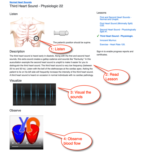
This diastolic murmurs module includes six lessons. We provide a textual description, audio recording, dynamic waveform video, and a cardiac animation for each lesson. Optionally, a quiz can be taken to measure comprehension and listening skills. Users who have selected our Auscultation-Basics, Essentials or Advanced plans can print achievement certificates and view their progress and scores using our personalized dashboard.
Overview
Diastolic murmurs are sustained sounds occurring between the second heart sound and first heart
sound of the next beat. These murmurs should be considered abnormal. Common sources of diastolic
murmurs include:
Stenosis of the mitral or tricuspid valves.
Aortic or pulmonic valve incompetence (regurgitation).
Increased blood flow through the mitral or tricuspid valves.
- Differentiate systolic from diastolic murmurs
- Identify murmur timing
- Recognize key features of the murmur's sound
- Gain exposure to abnormalities associated with diastolic murmurs
- Location, for example Erb's Point murmurs
- Timing: early, mid, late or pansystolic
- Loudness and its pattern during systole
- Pitch
- Quality (harsh, blowing or musical)
- Murmur changes during the respiratory cycle
The lessons in this course present several diastolic murmurs, their attributes and clinical correlations.
Start Diastolic Murmurs Course
Diastolic Murmurs Lessons
Introduction
Mild Aortic Regurgitation
In mild aortic regurgitation murmurs, the first heart sound is diminished due to premature closure of the mitral valve leaflets. An aortic ejection click follows the first heart sound by 75 milliseconds and the second heart sound is normal. Systole is silent. A high-pitched decrescendo murmur occupying the first half of diastole can be heard beginning immediately after the second heart sound.
This murmur should be auscultated at Erb's Point and can be accentuated by having the patient sitting up and leaning forward holding his breath after expiration.
The murmur can be caused by a bicuspid (thickened) aortic valve. In the cardiac animation, observe turbulent blood flow from the aorta into the left ventricle in early diastole. Also observe the minimally thickened aortic valve leaflets.
Mild Aortic Regurgitation Lesson
Mild Pulmonic Regurgitation
This lesson presents mild pulmonic regurgitation. Note that first and second heart sounds are normal (S2 is split) in the recording. Systole is silent. A high-pitched decrescendo murmur occurs in the first half of diastole. The murmur begins immediately after S2. The intensity of the murmur increases with inspiration, indicating a right-sided origin of the murmur.
Auscultate this murmur at the pulmonic area and with the patient sitting up and leaning forward.
In the animation you can see the turbulent blood flow from the pulmonary artery into the right ventricle during early diastole. You can see the minimally thickened pulmonic valve leaflets. This murmur can be caused by an infection of the pulmonic valve leaflets.
Mild Pulmonic Regurgitation Lesson
Mild Mitral Stenosis
Mild mitral stenosis murmurs can have increased intensity of S1, while the second heart sound is normal and unsplit. Systole is silent. In this recording, an opening snap occurs 100 milliseconds into diastole. As mitral stenosis becomes more severe, the opening snap will occur earlier in diastole. A diamond-shaped, low-frequency murmur follows the opening snap.
Auscultate using the stethoscope bell.
The first heart sound intensity is increased because of the mild thickening of the mitral valve leaflets. Mild mitral stenosis is commonly due to rheumatic heart disease.
In the cardiac animation, observe the turbulent blood flow from the left atrium into the left ventricle. Also, notice the minimally thickened mitral valve leaflets and the minimally enlarged left atrium.
Minimally Split First Heart Sound Lesson
Moderate Mitral Stenosis
This is an example of moderate mitral stenosis. The first heart sound is increased in intensity. The second heart sound is normal and unsplit. Systole is silent. There is an opening snap 75 milliseconds into diastole. As mitral stenosis becomes more severe, this opening snap will occur earlier in diastole.
A diamond-shaped, low-frequency murmur follows the opening snap. There is a second murmur in late diastole due to contraction of the left atrium.
Auscultate using the bell of the stethoscope.
Mitral stenosis can be caused by rheumatic heart disease, and this causes a thickening of the mitral valve leaflets. In the cardiac animation, observe turbulent blood flow from the left atrium into the left ventricle. Take note of the moderately thickened mitral valve leaflets and the moderately enlarged left atrium. The excursion of the mitral valve leaflets is moderately decreased.
Moderate Mitral Stenosis Lesson
Severe Mitral Stenosis
In this example of severe mitral stenosis, notice that the first heart sound has decreased intensity due to severe thickening of the mitral valve leaflets. The second heart sound is normal and unsplit, and systole is silent. An opening snap occurs 50 milliseconds into diastole. As mitral stenosis becomes more severe, the opening snap will occur earlier in diastole. The opening snap is followed by a low-frequency murmur which occupies the remainder of diastole. The first two-thirds of the murmur is diamond-shaped and the remainder is a crescendo.
Use the bell of the stethoscope to hear this murmur.
In the cardiac animation video, observe the turbulent blood flow from the left atrium into the left ventricle. Also notice the severely thickened mitral valve leaflets and the markedly enlarged left atrium. The excursion of the mitral valve leaflets is severely reduced. Mitral stenosis is commonly caused by rheumatic heart disease.
Moderate Tricuspid Stenosis
This lesson presents moderate tricuspid stenosis. The first heart sound has increased intensity due to moderate thickening of the tricuspid valve leaflets. The second heart sound is normal and unsplit. Systole is silent.
This murmur includes a tricuspid opening snap which is followed by a diamond-shaped low-frequency murmur. There is a second murmur in late diastole due to the right atrium contraction. For this condition, the murmur intensity and tricuspid opening snap increase with inspiration.
Auscultate with the bell of the stethoscope.
In the cardiac animation, observe the turbulent blood flow from the right atrium into the right ventricle. Also observe the moderately thickened tricuspid valve leaflets and the moderately enlarged right atrium. The excursion of the tricuspid valve leaflets is moderately decreased. Moderate tricuspid stenosis is most commonly due to rheumatic heart disease.
Moderate Tricuspid Stenosis Lesson
Course Quiz
After completing all lessons in a course, a quiz becomes available. If the user successfully completes a quiz, results are saved to the user's dashboard and a certificate can be printed.
Reference Guide
For subscribers, we provide a comprehensive heart sounds and murmurs reference guide. For each abnormality, one or more sound recordings are available along with text, phonocardiogram and cardiac animation.How To Use This Course

Authors and Reviewers
-
Heart sounds by Dr. Jonathan Keroes, MD and David Lieberman, Developer, Virtual Cardiac Patient.
- Reviewed by Dr. Barbara Erickson, PhD, RN, CCRN.
-
Last Update: 11/9/2021
Sources
-
Heart Sounds and Murmurs Across the Lifespan (with CD)
Dr Barbara Ann Erickson
Publisher: Mosby
ISBN-10: 0323020453; ISBN-13: 978-0323020459 -
Heart Sounds and Murmurs: A Practical Guide with Audio CD-ROM 3rd Edition
Elsevier-Health Sciences Division
Barbara A. Erickson, PhD, RN, CCRN - How to measure blood pressure using a manual monitor
Mayo Foundation for Medical Education and Research (MFMER) -
Heart and Lung Sounds Reference Guide
PracticalClinicalSkills.com -
Manual Blood Pressure Measurement
Vital Sign Measurement Across the Lifespan