Cardiac Procedures
Pericardiocentesis
Mayo Clinic experience: “Ideal entry” point in 211 cases

- 173 para-apical
- 12 left parasternal
- 12 left axillary
- 11 right parasternal
- 3 posterolateral
- Subcostal in 29 cases
parasternal
subxiphoid
Narration
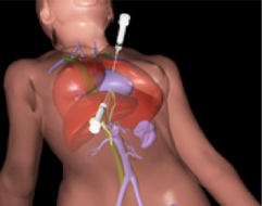
So pericardiocentesis is hopefully an infrequent procedure in the emergency setting, but it can certainly be aided with ultrasound guidance. Typically, it is taught to do this procedure blindly from a subxyphoid approach, but the thing to understand with this is getting to the pericardial sac through the liver can involve going through a lot of tissue and what has been found when you use ultrasound guidance you can find places where the skin is close to that pericardial fluid collection and in a large series from the Mayo Clinic they found that the majority of cases - over 80% - where in the periapical approach next to the apex of the heart they found a very short distance from skin to effusion and this was the preferred access route.
Pericardiocentesis
Narration
You can access the pericardium through a parasternal view and look at it from a subxyphoid view or vice versa. This shows a patient in a code situation where we have accessed the pericardial space anteriorly, the heart is not beating obviously, but the needle is in the pericardial space trying to aspirate that fluid.
Pericardiocentesis
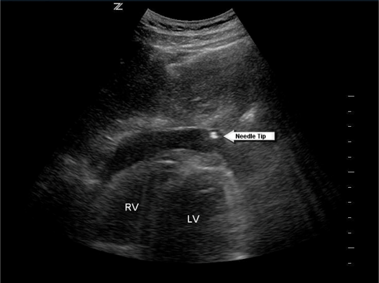
Can visualize subxiphoid with para-apical approach

Narration
Here is a still image showing a subxyphoid view of the heart where we are looking through the liver to see that pericardial tip and the needle was put in through a periapical approach, but we're visualizing it to make sure it is in the right place using the ultrasound probe in a different position.
Condition?
Narration
We can also use ultrasound guidance for other cardiac procedures. You look at this case, we'll talk about this more in the cardiac lecture, but this is a parasternal long axis view in the emergency medicine orientation, what you can see here is you've got atrial contraction, that movement of the valves going along at a regular rate and then you've got ventricular contraction, which is quite bradycardic. This is actually third degree heart block or complete heart block and the treatment for this is a pacer, which with ultrasound guidance you could do transvenously.
Pacer Placement And Capture
- Ultrasound guided central access
- Pacer through RA -> RV
- Capture
Narration
What you want to do there is obviously use ultrasound guidance for your central venous access and thread that catheter down and make sure it’s passing through the tricuspid into the tip of the right ventricle as seen here and you can also tell that there is capture, hopefully there will be a pulse as well, but ultrasound can be very helpful in guiding the various parts of this procedure.