More Pleural Effusions
Another Large Pleural Effusion

Narration
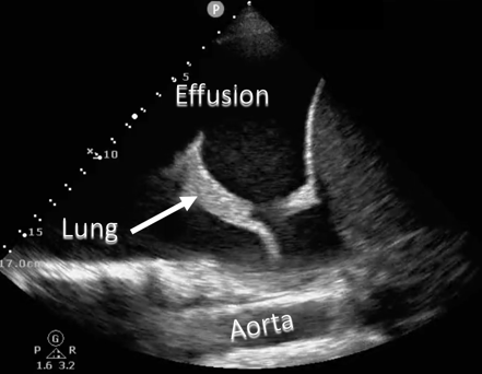
So here’s an example of another large right pleural effusion - you can see the fluid above the diaphragm and some lung that is again collapsed and sort of waving around in that pleural space, it’s well delineated from the fluid and actually in the far field of this image you can see the aorta, which you normally wouldn’t see except you have the fluid to look through.
Another Large Pleural Effusion


Narration
Here’s a labeled image that shows the effusion again above the diaphragm with the aorta in the far field continuing up behind the effusion.
Left Pleural Effusion

Narration
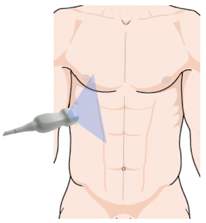
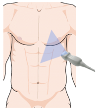
We can also of course look on the left for a pleural effusion. The image looks very similar, we simply place the probe again, the phased array probe in this case, on the left flank with the indicator towards the head and we can see the spleen and the kidney with the diaphragm above the spleen and again that black wedged shaped area that indicates a pleural effusion and we can also see the spine sign on the left.
Small Pleural Effusion

Narration
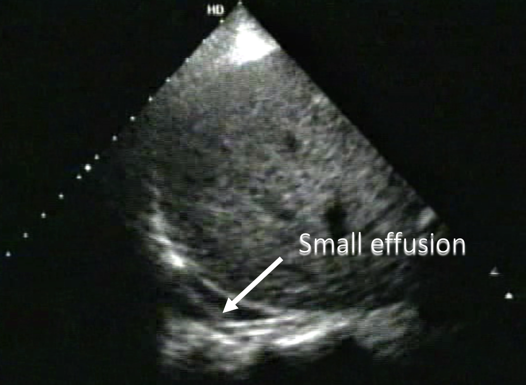
Here is an example of a small pleural effusion, this is on the right side you can see the liver. We’re not really seeing it anteriorly, but in the back you can again see that spine sign the very small triangular area of fluid with a little bit of lung waving around in it.
Small Pleural Effusion


Narration
This is that small effusion labeled in he still image along with the moving image up on the right.
Loculated Pleural Effusion

Narration
Occasionally you may see debris or loculations in the pleural effusion. This is typically a chronic process. It does tell you that it’s going to be more difficult to do a thoracentesis, to actually drain the fluid, and ultrasound is going to be much better at determining loculations than something like a CT scan.