Pneumonia
Pneumonia
- Lung consolidation
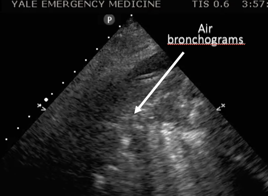
- "Air bronchograms"
Narration
So typically when we look at the chest, if the lung is well aerated, we really aren’t going to see the lung, we’ll see the lining of the lung the visceral pleura, but when there’s pathology - consolidation from something such as pneumonia you can actually sometimes see the lung tissue and within the lung tissue you may see areas of air which are going to be bright on US and these are what are called air bronchograms.
Pneumonia

Narration
So here’s a still image pointing out those bright hyperechoic air bronchograms within consolidated lung tissue, in this case also within a pleural effusion or parapneumonic effusion surrounding the consolidating lung.
Pneumonia
- Small consolidation
- Pediatric patient
Narration
You may be able to see consolidation using a linear probe, this was a pediatric patient that did have a pneumonia that was seen on US when interrogating the right posterior lung field.
Pneumonia
- “Hepatization”
- Lung looks like liver
Narration
Occasionally when the lung is completely socked in and consolidated it may actually appear like the liver and this is what is called hepatization. In this image you can see consolidated lung above the diaphragm that looks a lot like the liver, and this is a bad pneumonia.