Morison's Pouch
Introduction

Narration
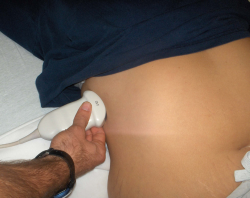
So this image is showing Morison’s pouch or the hepatorenal space - this is a key part of the fast examination, it’s the most sensitive area for fluid in the peritoneum. It can come from trauma but also from medical causes like ascites or ruptured ectopic or cyst. We’re using a curvilinear probe placed in the right flank or coronal view with the indicator towards the head.
Normal Morison’s Pouch - Hepatorenal Space


Narration
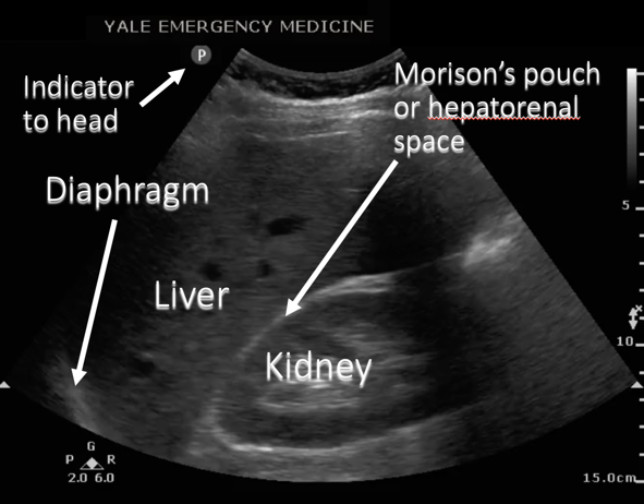
This is a schematic showing the liver, the kidney, and that space between the two which is Morison’s Pouch, the diaphragm is to the left of the screen or superior with the indicator towards the patients head.
Large Free Fluid In Morison’s Pouch

Narration
So in this clip we see a large amount of fluid, this was actually a ruptured ectopic, there’s some clots starting to form. This probably represents more than a liter-liter and a half of blood in the peritoneum, with a large amount of fluid (black area) between the liver and the kidney.
Small Free Fluid In Morison’s Pouch

Narration
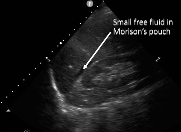
This clip on the other hand shows a small amount of fluid in Morrisons pouch, probably around the order of 500ccs maybe a little bit less, which is about the borderline where you’re going to be very sensitive for finding fluid. But you can see that black stripe between the liver and the kidney and it really just kind of tracks into crevasses in that space.
Small Free Fluid In Morison’s Pouch


Narration
This is a diagram showing that small amount of free fluid which is collecting in Morison’s pouch between the liver and the kidney.