Pelvic Free Fluid
Free Fluid In Pelvis - Transverse
Narration
So this is transverse view of the pelvis showing a large amount of free fluid and it is surrounding the uterus, in this case in the female pelvis.
Free Fluid In Pelvis - Transverse

Narration
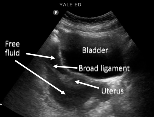
This is a schematic we can see the uterus and the broad ligament with free fluid both anterior and posterior in addition to fluid within the bladder that is anterior and well contained within the bladder wall.
Free Fluid In Pelvis - Sagittal

Narration
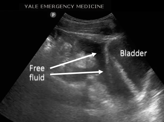
We can rotate the image into a sagittal plane with the indicator towards the head and the bladder is seen here on the right side of the screen and this is particularly helpful in the male pelvis where you don’t have the uterus to sort of help delineate the fluid.
Free Fluid In Pelvis - Sagittal

Narration
Diagram shows the bladder with the contained fluid and the free fluid superior to the bladder.
Free Fluid In Pelvis - Sagittal

Narration
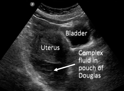
In the female pelvis you should be able to see the uterus and this is a sagittal image again with the bladder anteriorly, the uterus is superior to the bladder and there is a large amount of complex fluid both anterior and posterior to the uterus. You can see some echoes within it and again this is a large amount of fluid probably around the order of a liter, and I believe this was a ruptured ectopic pregnancy.
Free Fluid In Pelvis - Sagittal

Narration
You can see that complex fluid in the rectouterine pouch, also known as the pouch of Douglas, along with the bladder with the contained fluid anteriorly.
Free Fluid In Pelvis - Transverse
Narration
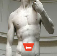
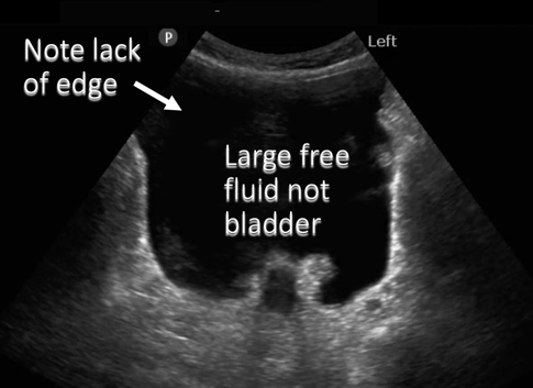
So here’s an examination in the pelvis, and it’s transverse, and it actually looks a bit like the bladder when you first look at it, but you need to be careful here because if you track this fluid you can't actually see the bladder wall up towards the superior corners.
Free Fluid In Pelvis - Transverse

Narration
And this is actually large free fluid in the pelvis with the aorta in the far field and you’ll note that there is no encapsulation of this fluid, there’s no bladder wall keeping it in its place.