Fluid In & Around Kidney
Fluid Within The Kidney - Hydro

Narration
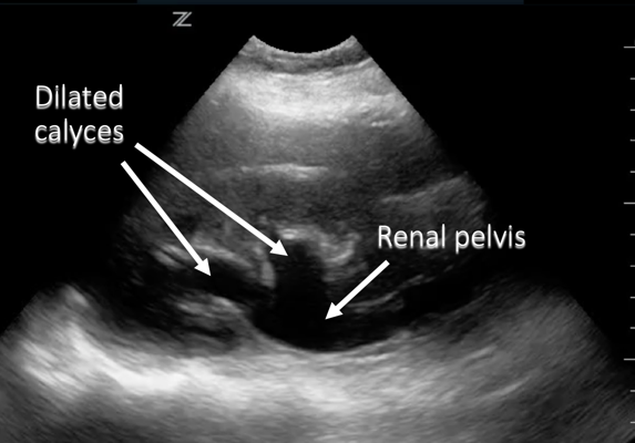
So another place we can look for fluid again from the right upper quadrant or flank view, same view we use for Morison’s pouch, but actually interrogating the kidney for fluid within the kidney, and this is called hydronephrosis when we have fluid that is backing up into the calyces and down into the renal pelvis.
Fluid Within The Kidney - Hydro


Narration
Diagram showing those dilated calices, the renal pelvis, this is most commonly caused by a kidney stone that is going to block the ureter and back up the fluid. In this case this is probably moderate hydro, well talk about that more in the renal lecture, but it can be graded as mild, moderate, or severe hydro. You do need to watch out for the vasculature and sometimes color flow can help delineate that.
Fluid In And Around The Kidney

Narration
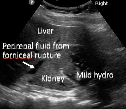
In addition to some hydronephrosis within the kidney, occasionally you’ll get fluid that ruptures outside of the kidney but is actually within the renal capsule and not within Morisons pouch. So here we see some fluid around the kidney and this is actually a forniceal rupture or a calyx that has ruptured you can see the kidney stones and some mild hydronephrosis here.
Fluid In And Around The Kidney – Mild Hydronephrosis With Forniceal Rupture


Narration
This is a diagram showing the liver, the perirenal fluid from the forniceal rupture, the kidney with the mild hydro and some kidney stones in it.