Sagittal
Rotating Transverse To Sagittal

Narration
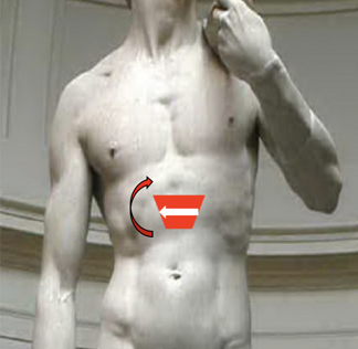
Once we have a good transverse view of the aorta we can rotate into a sagittal view, keeping the aorta in view. I like to do this to make sure we’re actually visualizing the aorta and not the IVC. Here’s the rotation clockwise, so the indicator goes towards the patient’s head and you can see that sagittal view of the aorta.
Sagittal Aorta

Narration
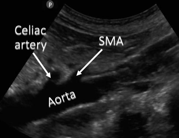
When you get a good sagittal view you can see the two proximal branches of the aorta, the celiac artery and the superior mesentery artery. Very occasionally you might see the inferior mesentery artery, but typically it’s the celiac and SMA that are most well visualized.
Sagittal Aorta


Narration
Here’s a cut away labeled with that first branch of the abdominal aorta, the celiac artery, coming off just above the SMA, you see the aorta sitting on those vertebral bodies which comes closer to the abdominal wall as it goes distally