Measurements
AAA Transverse

Narration
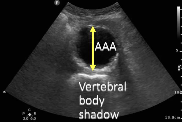
When we measure the aorta, we typically want to do it in a transverse view and we want to go from outside wall to outside wall to make sure we are not underestimating any aneurysm and we are visualizing the entire aorta. This shows a transverse view of a AAA probably between 4 and 5 cm and it would be measured again from the tips of these arrows which would be outside wall to outside wall.
AAA Sagital

Narration
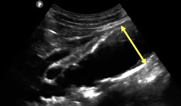
We can also see an aneurysm if we rotate into a sagittal view; in this case there is an aneurysm probably about 4 cm. You can see the proximal branches of the aorta and then the infarenal AAA (or abdominal aortic aneurysm) clot or thrombus in the actual wall of the aorta
AAA Sagittal

Narration
And if we were to measure this, again we would do it perpendicular to the axis of the lumen and include the entire aorta from outside to outside.
AAA Measure: Outside To Outside

Narration
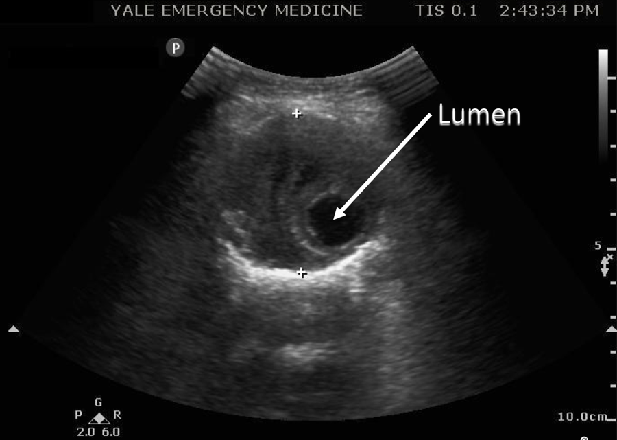
Here’s an example of that measurement, in this case with the lumen shone where the actual blood is traveling, but the aneurysm includes the entire area between those two cursors.
AAA Measure

Narration
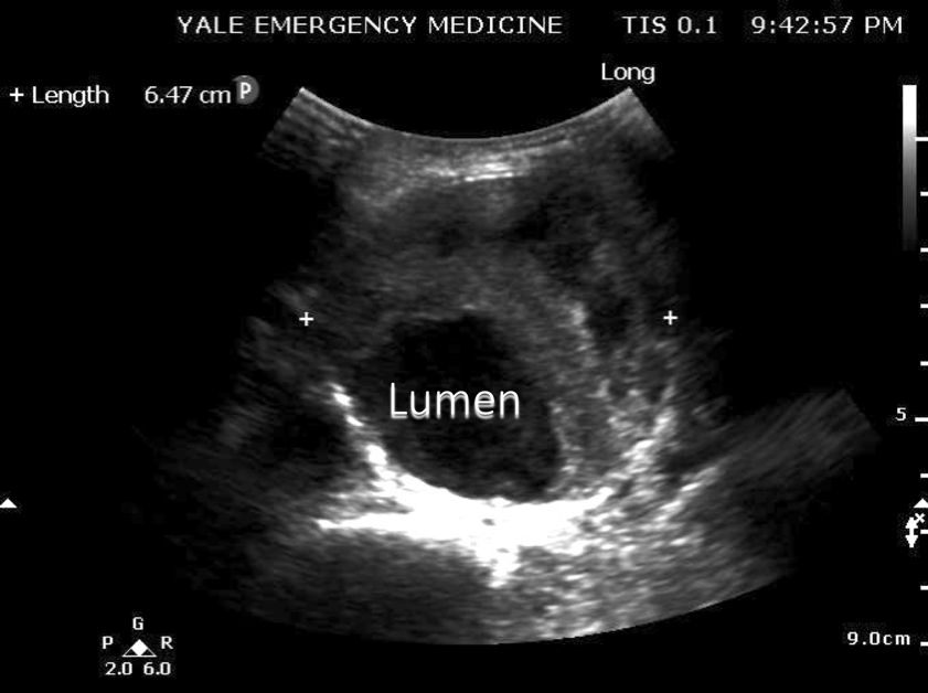
Here’s another AAA, measured in this cased from side to side, at about 6.5 cm with atherosclerosis, or clot, seen outside of the true lumen of the abdominal aorta.
AAA Measure

Narration
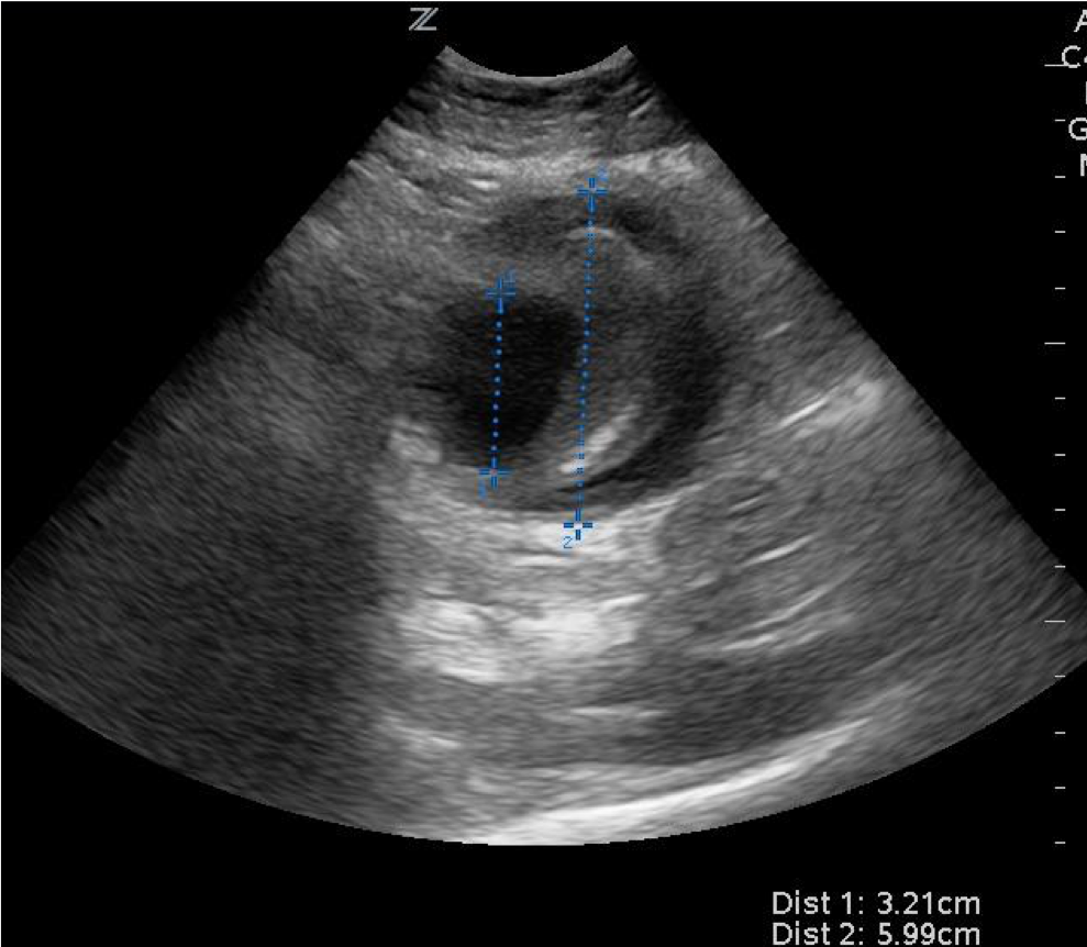
Here’s another one in a transverse view showing the lumen of the aorta measuring at 3.2 but the entire aneurysm close to 6 cm with a hypoechoic sort of crescented black area that probably does represent some leaking of the AAA.