IVC Abnormalities
IVC Pathology

Narration
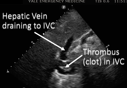
So we’re primarily going to use the IVC to assess fluid or hydration status from a flat collapsible IVC that could benefit from hydration versus a dilated non collapsible IVC that may be present in fluid overload or CHF. On occasion we can see other pathologies such as this IVC where we can see a hyperechoic area waving around there that is actually a clot or thrombus.
IVC Pathology


Narration
Here's a diagram showing that thrombus in transit, it could actually benefit from lytic therapy or interventional radiology to get it out.
IVC Pathology

Narration
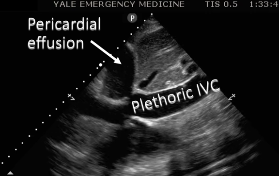
The other things the IVC can be helpful with are assessing for tamponade physiology. Here we see an image of a sagittal view of the IVC in the inferior pericardium and there’s a moderate to large pericardial effusion (fluid collection around the heart) that’s preventing filling into the heart and therefore the IVC is dilated and this indicates…
IVC Pathology


Narration
Tamponade physiology. Here’s a diagram showing that plethoric or dilated IVC with a large pericardial effusion.